Table of Contents


Cell — Structure and Functions
You have already learnt that things around us are either living or non-living. Further, you may recall that all living organisms carry out certain basic functions. Can you list these functions?
Different sets of organs perform the various functions you have listed. In this chapter, you shall learn about the basic structural unit of an organ, which is the cell. Cells may be compared to bricks. Bricks are assembled to make a building. Similarly, cells are assembled to make the body of every organism.
8.1 Discovery of the Cell
Robert Hooke in 1665 observed slices of cork under a simple magnifying device. Cork is a part of the bark of a tree. He took thin slices of cork and observed them under a microscope. He noticed partitioned boxes or compartments in the cork slice (Fig. 8.1).
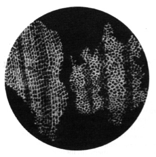
These boxes appeared like a honey- comb. He also noticed that one box was separated from the other by a wall or partition. Hooke coined the term ‘cell’ for each box. What Hooke observed as boxes or cells in the cork were actually dead cells.
Cells of living organisms could be observed only after the discovery of improved microscopes. Very little was known about the cell for the next 150 years after Robert Hooke’s observations. Today, we know a lot about cell structure and its functions because of improved microscopes having high magnification.
8.2 The Cell
Both, bricks in a building and cells in the living organisms, are basic structural units [Fig. 8.2(a), (b)]. The buildings, though built of similar bricks, have different designs, shapes and sizes. Similarly, in the living world, organisms differ from one another but all are made up of cells. Cells in the living organisms are complex living structures unlike non-living bricks.
A hen’s egg can be seen easily. Is it a cell or a group of cells?

The egg of a hen represents a single cell and is big enough to be seen by the unaided eye.
8.3 Organisms show Variety in Cell Number, Shape and Size
How do scientists observe and study the living cells? They use microscopes which magnify objects. Stains (dyes) are used to colour parts of the cell to study the detailed structure.
There are millions of living organisms. They are of different shapes and sizes. Their organs also vary in shape, size and number of cells. Let us study about some of them.
Number of Cells
Can you guess the number of cells in a tall tree or in a huge animal like the elephant? The number runs into billions and trillions. Human body has trillions of cells which vary in shapes and sizes. Different groups of cells perform a variety of functions.
A billion is a thousand million. A trillion is a thousand billion.
Organisms made of more than one cell are called multicellular (multi : many; cellular : cell) organisms. The number of cells being less in smaller organisms does not, in any way, affect the functioning of the organisms. You will be surprised to know that an organism with billions of cells begins life as a single cell which is the fertilised egg. The fertilised egg cell multiplies and the number of cells increase as development proceeds.
Look at Fig 8.3 (a) and (b). Both organisms are made up of a single cell. The single-celled organisms are called unicellular (uni : one; cellular : cell) organisms.
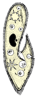
A single-celled organism performs all the necessary functions that multicellular organisms perform.
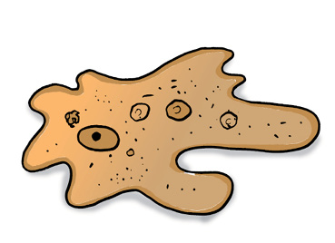
A single-celled organism, like amoeba, captures and digests food, respires, excretes, grows and reproduces. Similar functions in multicellular organisms are carried out by groups of specialised cells forming different tissues. Tissues, in turn, form organs.
Activity 8.1
The teacher may show a permanent slide of amoeba and paramecium under a microscope. Alternatively, the teacher can collect pond water and show these organisms by preparing the slides.
Shape of Cells
Refer to Fig. 8.3 (a). How do you define the shape of amoeba in the figure? You may say that the shape appears irregular. Infact, amoeba has no definite shape, unlike other organisms. It keeps on changing its shape. Observe the projections of varying lengths protruding out of its body. These are called pseudopodia (pseudo : false; podia : feet), as you learnt in Class VII. These projections appear and disappear as amoeba moves or feeds.
What advantage does amoeba derive by changing shape?
The change in shape is due to formation of pseudopodia which facilitates movement and help in capturing food.
A white blood cell (WBC) in human blood is another example of a single cell which can change its shape. But while WBC is a cell, amoeba is a full fledged organism capable of independent existence.
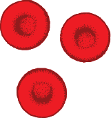
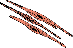
What shape would you expect in organisms with millions of cells? Fig. 8.4 (a, b, c) shows different cells such as blood, muscle and nerve of human beings. The different shapes are related to their specific functions.
Generally, cells are round, spherical or elongated [Fig. 8.4(a)]. Some cells are long and pointed at both ends. They exhibit a spindle shape [Fig. 8.4(b)]. Cells sometimes are quite long. Some are branched like the nerve cell or a neuron [Fig. 8.4(c)]. The nerve cell receives and transfers messages, thereby helping to control and coordinate the working of different parts of the body.
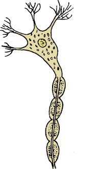
(c)
Can you guess, which part of the cell gives it shape? Components of the cell are enclosed in a membrane. This membrane provides shape to the cells of plants and animals. Cell wall is an additional covering over the cell membrane in plant cells. It gives shape and rigidity to these cells (Fig. 8.7). Bacterial cell also has a cell wall.
Size of Cells
The size of cells in living organisms may be as small as a millionth of a metre (micrometre or micron) or may be as large as a few centimetres. However, most of the cells are microscopic in size and are not visible to the unaided eye. They need to be enlarged or magnified by a microscope. The smallest cell is 0.1 to 0.5 micrometre in bacteria. The largest cell measuring 170 mm ×130 mm, is the egg of an ostrich.
Activity 8.2
Boil a hen’s egg. Remove the shell. What do you observe? A white material surrounds the yellow part. White material is albumin which solidifies on boiling. The yellow part is yolk. It is part of the single cell. You can observe this single cell without any magnifying device.
Are the cells in an elephant larger than the cells in a rat?
The size of the cells has no relation with the size of the body of the animal or plant. It is not necessary that the cells in the elephant be much bigger than those in a rat. The size of the cell is related to its function. For example, nerve cells, both in the elephant and rat, are long and branched. They perform the same function, that of transferring messages.
8.4 Cell Structure and Function
You have learnt that each living organism has many organs. You have studied in Class VII about the digestive organs which together constitute the digestive system. Each organ in the system performs different functions such as digestion, assimilation and absorption. Similarly, different organs of a plant perform specific/specialised functions. For example, roots help in the absorption of water and minerals. Leaves, as you have learnt in Class VII, are responsible for synthesis of food.
Each organ is further made up of smaller parts called tissues. A tissue is a group of similar cells performing a specific function.
Paheli realised that an organ is made up of tissues which in turn, are made up of cells. The cell in a living organism is the basic structural unit.
8.5 Parts of the Cell
Cell Membrane
The basic components of a cell are cell membrane, cytoplasm and nucleus (Fig. 8.7). The cytoplasm and nucleus are enclosed within the cell membrane, also called the plasma membrane. The membrane separates cells from one another and also the cell from the surrounding medium. The plasma membrane is porous and allows the movement of substances or materials both inward and outward.
Activity 8.3
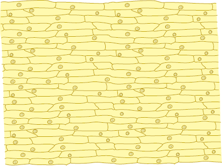
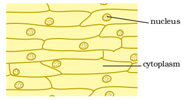
In order to observe the basic components of the cell, take an onion bulb. Remove the dry pink coverings (peels). You can easily separate these from the fleshy white layers of the bulb with the help of forceps or even with your hand. You can also break the onion bulb and separate out thin layers. Place a small piece of the thin onion peel in a drop of water on a glass slide. The thin layer can be cut into smaller pieces with the help of a blade or forceps. Add a drop of methylene blue solution to the layer and place a coverslip on it. While placing the coverslip ensure that there are no air bubbles under the coverslip. Observe the slide under the microscope. Draw and label. You may compare it with Fig. 8.5.
The boundary of the onion cell is the cell membrane covered by another thick covering called the cell wall. The central dense round body in the centre is called the nucleus. The jelly-like substance between the nucleus and the cell membrane is called cytoplasm.
You have learnt earlier that the cell membrane gives shape to the cell. In addition to the cell membrane, there is an outer thick layer in cells of plants called cell wall. This additional layer surrounding the cell membrane is required by plants for protection. Plant cells need protection against variations in temperature, high wind speed, atmospheric moisture etc. They are exposed to these variations because they cannot move. Cells can be observed in the leaf peel of Tradescantia, Elodea or Rhoeo. You can prepare a slide as in the case of onion.
Paheli asks Boojho if he can also observe animal cells.
Activity 8.4
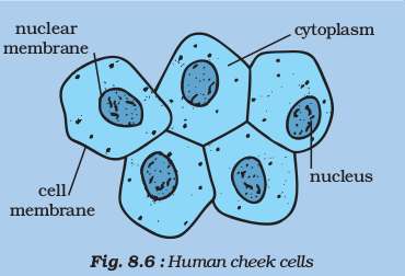
Take a clean tooth pick, or a matchstick with the tip broken. Scrape inside of your cheek without hurting it. Place it in a drop of water on a glass slide. Add a drop of iodine and place a coverslip over it. Alternatively, add 1-2 drops of methylene blue solution. Observe it under the microscope. You may notice several cells in the scraped material (Fig. 8.6). You can identify the cell membrane, the cytoplasm and nucleus. A cell wall is absent in animal cells.
Cytoplasm
It is the jelly-like substance present between the cell membrane and the nucleus. Various other components, or organelles, of cells are present in the cytoplasm. These are mitochondria, golgi bodies, ribosomes, etc. You will learn about them in later classes.
Nucleus
It is an important component of the living cell. It is generally spherical and located in the centre of the cell. It can be stained and seen easily with the help of a microscope. Nucleus is separated from the cytoplasm by a membrane called the nuclear membrane. This membrane is also porous and allows the movement of materials between the cytoplasm and the inside of the nucleus.
With a microscope of higher magnification, we can see a smaller spherical body in the nucleus. It is called the nucleolus. In addition, nucleus contains thread-like structures called chromosomes. These carry genes and help in inheritance or transfer of characters from the parents to the offspring. The chromosomes can be seen only when the cell divides.
Gene
Gene is a unit of inheritance in living organisms. It controls the transfer of a hereditary characteristic from parents to offspring. This means that your parents pass some of their characteristics on to you. If your father has brown eyes, you may also have brown eyes. If your mother has curly hair, you might also end up having curly hair. However, the different combination of genes from parents result in different characteristics.
Nucleus, in addition to its role in inheritance, acts as control centre of the activities of the cell. The entire content of a living cell is known as protoplasm. It includes the cytoplasm and the nucleus. Protoplasm is called the living substance of the cell.
Paheli wants to know if the structure of the nucleus is the same in cells of plants, animals and bacteria.
The nucleus of the bacterial cell is not well-organised like the cells of multicellular organisms. There is no nuclear membrane. The cells having nuclearmaterial without nuclear membrane are termed prokaryotic cells. The organismswith these kinds of cells are called prokaryotes (pro : primitive; karyon : nucleus). Examples are bacteria and blue green algae. The cells, like onion cells and cheek cells having well-organised nucleus with a nuclear membrane are designated as eukaryotic cells. All organisms other than bacteria and blue green algae are called eukaryotes. (eu : true; karyon: nucleus).
While observing the onion cells under the microscope, did you notice any blank-looking structures in the cytoplasm? It is called vacuole. It could be single and big as in an onion cell. Cheek cells have smaller vacuoles. Large vacuoles are common in plant cells. Vacuoles in animal cells are much smaller.
You might have noticed several small coloured bodies in the cytoplasm of the cells ofTradescantia leaf. They are scattered in the cytoplasm of the leaf cells. These are called plastids. They are of different colours. Some of them contain green pigment called chlorophyll. Green coloured plastids are called chloroplasts. They provide green colour to the leaves. You may recall that chlorophyll in the chloroplasts of leaves, is essential for photosynthesis.
8.6 Comparison of Plant and Animal Cells
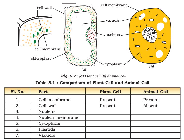
If you recall Activities 8.3 and 8.4, you should be able to compare plant and animal cells. Observe the plant and animal cell carefully in Fig. 8.7 (a), (b).
Let us tabulate the similarities and disinguishing features of plant and animal cells. Only a few features are mentioned. You may add more in
Table 8.1.
.
Keywords
Cell
Cell membrane
Cell wall
chloroplast
chromosome
Cytoplasm
Eukaryotes
gene
Multicellular
Nuclear membrane
Nucleolus
Nucleus
Organ
Organelles
plasma membrane
plastid
Prokaryotes
Pseudopodia
Tissue
Unicellular
vacuole
White blood cell (WBC)
What you have learnt
- All organisms are made of smaller parts called organs.
- Organs are made of still smaller parts. The smallest living part of an organism is a ‘cell’.
- Cells were first observed in cork by Robert Hooke in 1665.
- Cells exhibit a variety of shapes and sizes.
- Number of cells also varies from organism to organism.
- Some cells are big enough to be seen with the unaided eye. Hen’s egg isan example.
- Some organisms are single-celled, while others contain large number of cells.
- The single cell of unicellular organisms performs all the basic functions performed by a variety of cells in multicellular organisms.
- The cell has three main parts: (i) the cell
- membrane, (ii) cytoplasm which contains smaller components called organelles, and
- (iii) the nucleus.
- Nucleus is separated from cytoplasm by a nuclear membrane.
- Cells without well-organised nucleus, i.e.
- lacking nuclear membrane, are called prokaryotic cells.
- Plant cells differ from animal cells in having an additional layer around the cell membrane termed cell wall.
- Coloured bodies called plastids are found in the plant cells only. Green plastids containing chlorophyll are called chloroplasts.
- Plant cell has a big central vacuole unlike a number of small vacuoles in animal cells.
Exercises
1. Indicate whether the following statements are True (T) or False (F).
(a) Unicellular organisms have one-celled body. (T/F)
(b) Muscle cells are branched. (T/F)
(c) The basic living unit of an organism is an organ. (T/F)
(d) Amoeba has irregular shape. (T/F)
2. Make a sketch of the human nerve cell. What function do nerve
cells perform?.
3. Write short notes on the following.
(a) Cytoplasm
(b) Nucleus of a cell
4. Which part of the cell contains organelles?
5. Make sketches of animal and plant cells. State three differences
between them.
6. State the difference between eukaryotes and prokaryotes.
7. Where are chromosomes found in a cell? State their function.
8. ‘Cells are the basic structural units of living organisms’. Explain.
9. Explain why chloroplasts are found only in plant cells?
10. Complete the crossword with the help of clues given below.
Across
1. This is necessary photosynthesis.
3. Term for component present in the cytoplasm.
6. The living substance in the cell.
8. for Units of inheritance present on the chromosomes.
Down
1. Green plastids.
2. Formed by collection of tissues.
4. It separates the contents of the cell from the surrounding medium.
5. Empty structure in the cytoplasm.
7. A group of cells.
Extended Learning — Activities and Projects
1. Visit a laboratory for senior secondary students in your school or in a neighbouring school. Learn about the functioning of a microscope in the laboratory. Also observe how a slide is observed
under the microscope.
2. Talk to the senior biology teacher in your school or a neighbouring school. Find out if there are diseases which are passed on from parents to the offspring. Find out how these are carried and also if these diseases can be treated. For this you can also visit a doctor.
3. Visit an agriculture extension centre in your area. Find out about genetically modified (GM) crops. Prepare a short speech for your class on this topic.
4. Find out about Bt cotton from an agriculture expert. Prepare a short note on its advantages/disadvantages.
Did You Know?
The cells in the outermost layer of our skin are dead. An average adult carries around about 2 kg of dead skin. Billions of tiny fragments of the skin are lost every day. Every time you run your finger on a dusty table, you shed a lot of old skin.